
This week’s case comes from a fascinating share by a colleague, reminding us all why we should never ignore an automated analyser flag!
The 52 year old patient presented to the emergency department with an unexplained fever. The initial FBC showed a normal white cell count but a closer look at the parameters revealed a deeper issue.
Key FBC Results:
-
WBC: 4.99 x 10⁹/L
-
Hb: 124 g/L
-
Platelets: 58 x 109/L
The analyser flagged the sample for an abnormal platelet distribution. Looking at a platelet count of 58, the biomedical scientist on duty immediately performed a wet preparation to check for EDTA induced platelet clumping.
Instead of clumps, they noticed the following on the wet prep:
A blood film was made:
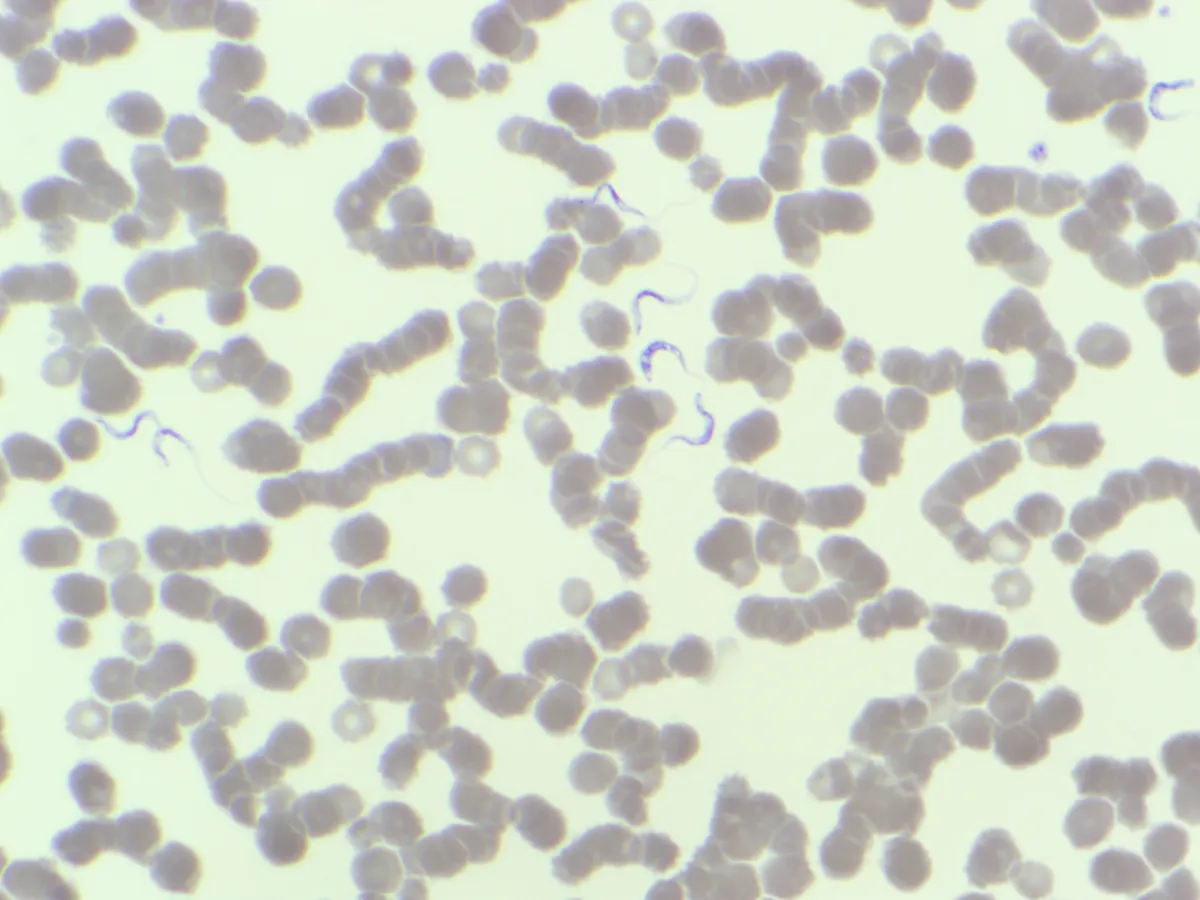
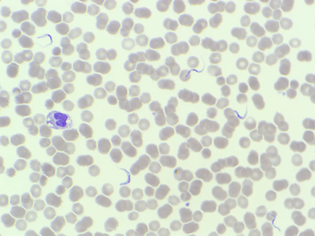
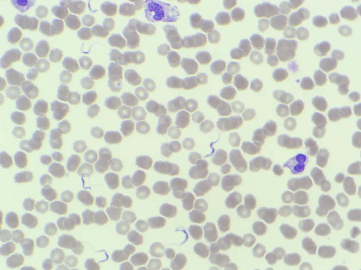
Drop your guesses and observations in the comments! As always, you can follow the discussion on LinkedIn.
Special thanks to Cha Wei Lee for sharing this case and blood film.